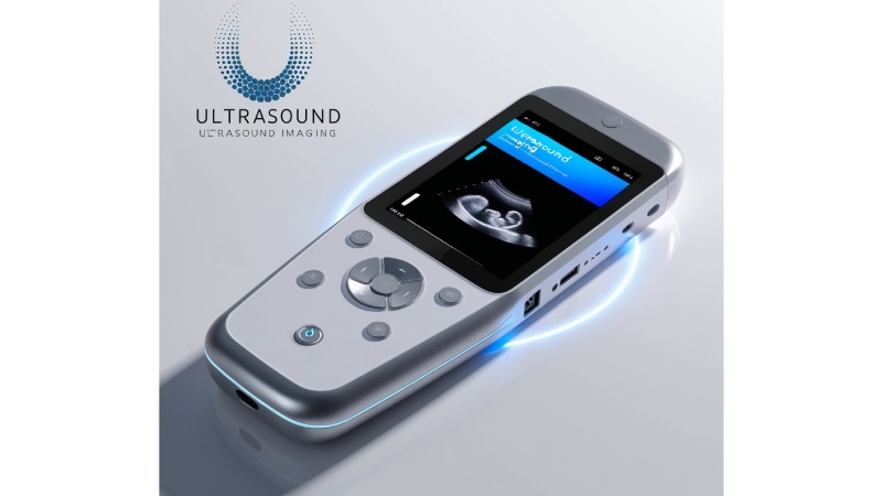
Portable compact size of the ultrasound machines has evolved in recent years to compact handheld machines. These small and portable, devices which use to be employed in large hospitals and particular clinics, are slowly finding their way to a myriad of health care institutions. Whether it’s in emergency departments or GP surgeries, on Mountains or in the bush, POC ultrasound is expanding the capacity for dx, mntnc & tretmnt at the pt of care. But how these machines are constructed, and being increasingly important in today’s medical practice?
What is ultrasound and how does it stand to work?
More basically, ultrasound is one of the types of the sound wave that help the practitioners to see through the body. Ultrasound is also different from x-ray, CT scans, which involves ionizing radiation to map the body parts but instead it uses sound waves. These sound waves are usually produced through a probe (or transducer) to be placed on the skin surface. As the sound waves penetrate the internal structures of the body they reflect off tissues and organs then back into the probe where they are amplified in the form of electrical signals. These signals are processed by the machine then it produces images which are viewed on a monitor.
The frequency of sound waves used in medical ultrasound is ordinarily in the range of 1 and 20MHz which is above the audible frequency range of 20Hz to 20kHz. The higher the frequency the better the resolution of the picture but the limit of penetration depth decreases with increase in frequency. As for various parts of the body, for body muscles, organs, or fetuses, different frequencies are used, so as to achieve the best resolution and penetration depth of the picture.
Ultrasound Portables’ history
Though the concept of ultrasound technology was first introduced in the 1950s, portable ultrasound devices that could be hand held has only become a reality recently. Portable and transportable ultrasound devices are considered possible solutions if only miniaturization and advances in transducers will happen. Most previous designs of ultrasound systems have been large, heavy, and professionally complex to operate. For example, these machines were also costly and thus were mostly a preserve of hospitals and large health facilities.
The first development strides in portable ultrasound emerged in the last decade of the 20th century and was characterized by the evolution of transducers small enough that they could be incorporated into portable machines without any compromise on image quality. There are many aspects that contributed to the development of today’s handheld units; including the developments in the digital signal processing (DSP)as well as the micro electronics. These changes made the development of compact ultrasound machines possible, in size, price, operating mechanisms and functionality.
Presently, handheld portable ultrasound machine have flexible dimensions and designs. Some are entirely portable, communicating with a portable device or computer, while others are integrated with an observer interface. It was also observed that these compact gadgets are capable of delivering fairly detailed images, so that diagnoses could be made at the place of contact.
What Makes a Handheld Ultrasound Machine Go?
A typical handheld ultrasound machine consists of several key components:
- Transducer: The transducer is the main component of the ultrasound machine. It is the source and the receiver of sound waves. Most of the handheld devices in use in the present world employ piezoelectric crystals that vary their shape depending on the electrical signals received. They produce the high frequency sounds waves when connected to electricity source and change the reflected echoes to electrical signals.
- Processing Unit: The processing unit is the computer in the device that draws out the received signals and converts it into useful image data. This unit uses sophisticated equations to increase image sharpness, detail and to remove unwanted artifacts.
- Display Screen: Standard ultrasound equipment include the large screen control panel while portable devices are equipped with small screens with very high pixel resolutions. These screens are of added advantage since it is possible for the health care provider to view the images on the device and thereby assess the results on the same screen.
- Power Source: Almost all the handheld ultrasound devices are operated through rechargeable lithium-ion batteries, and they can work for several hours at an instance. One aspect of battery-powered portability is one of the features that makes handheld devices different from the traditional devices that normally need an external power source.
- Wireless Connectivity: Some of the newer systems of handheld ultrasound systems are fit with Wi-Fi or Bluetooth which connects the device and the USG images to a mobile phone, tablet or the cloud. Such wireless capability allows for telemedicine, exchange of information, and linkages to EHR systems.
Imaging is one of the most common terms used in diagnosing an illness diagnosis and this write-up will attempt to look at the science behind the imaging.
In the case of ultrasound imaging, physics involves sound waves at different tissue types in the body. If ultrasound waves reach a boundary between tissues with different acoustical characteristics (muscle and fat or soft tissue and bone), part of the sound waves are reflected back to the transducer and the rest continue through. This differential reflection is used to ask a question, to answer a question, to express the idea, and to form an image.
In essence, the accuracy of the produced images is determined by the functionality of the transducer to transmit and to receive the sound waves properly. Scientists have come up with techniques on how to enhance quality of such images by especially for those portable equipments that lacks space. For example, digital beamforming is the method that enables the device to direct the sound waves toward the necessary depths, increase structural resolution on the deeper object parts, and keep the superficial object image clear.
This same technology is also used in handheld ultrasound devices to determine blood flow. Doppler ultrasound estimates the difference in the frequencies of the reflected sound waves relative to the moving structure (for example red blood corpuscles). When looking at this regard, types of blood flow can be assessed regarding pace and direction-which are applicable in conditions such as deep vein thrombosis or diseases involving the heart.
Significance of Portable Diagnostic Ultrasound Imaging
Portable ultrasound devices are making profound even in multiple specialties of medical practice. The attributes of mobility, simplicity and affordability have rendered them indispensable tools in emergency medicine, primary care, obstetrics and much more. Here are a few key reasons why handheld ultrasound machines matter:
1. Point-of-Care Diagnosis
Portable ultrasound scanners make diagnosis available at point-of-care and provide for decision-making at the point of need. For example, severe trauma or cardiopulmonary arrest, it can be valuable to do an extend head to toe ultrasound in a short amount of time to look for conditions such as hemorrhage or cardiac tamponade. He said this immediate feedback can inform the treatment process and may prevent more patients from dying.
2. Improved Access to Healthcare
It is very rare, especially in the rural or areas where there are few health facilities to have advanced diagnosis equipment. Handheld ultrasound machines are less expensive than conventional ultrasound machines and make imaging solutions accessible to areas where costs are prohibitive. This assist in early diagnosis of diseases and enhanced control of long-term illnesses.
3. Reduced Healthcare Costs
The small size and cost of the handheld ultrasound machines making them cheaper than the conventional ultrasound devices. The lack of expensive equipment and the availability of easily accessible internet from most clinics or practices means that these devices can be a significant diagnostic benefit for those offices that cannot afford multi-thousand dollar diagnostic tools. Also, by helping make diagnoses quicker, they can help cut out expensive referals and other tests.
4. Enhanced Patient Experience
Through portable ultrasound devices, chances of transports to imaging centers are minimized, waiting periods are also short, and results are presented quickly making patients happy. Patients receive valued care through an optimised and less demanding approach to receiving their care such as when the patient is able to view the images that their physician is using in consultation with them.
Revolutionary in the Field of Portable Ultrasound
As the handheld ultrasound devices , many people begin to see more and more, the practice is not cut off and ever-evolving research is still being made to improve the capability of these machines. Besides, some of the scientists have done tremendous work which includes Dr. Sanjiv Sam Gambhir, who has developed innovative ways of using ultrasound to check for early signs of cancer and other diseases, where they progress to to monitor diseases progress.
Some other researchers are also working on the use of integrating AI with handheld ultrasound machines. Due to the non-specialist nature of the technology, AI may improve the interpretative ability of the images to the extent that the end users do not necessarily have to be experts in the use of the invention. Such advances are making it easier and more natural for future handheld ultrasound to develop in the future.
Conclusion
Portable compact ultrasound devices can be singled out as one of the evidence that technological enhancements of medical instruments boost patient’s quality of lives in different countries. Since they offer instant, minimally invasive imaging at the patient-side, they are revolutionizing the strategy through which clinicians diagnose, manage, and treat various diseases. Looking into the future as these technologies are developed and further researched handheld ultrasound systems will be used more broadly to increase access to healthcare, increase successful diagnoses and treatment, as well as decrease costs.
Author Bio:
James Brown is also an innovative healthcare solutions enthusiast and the co-founder of BeWellFinder. James, who has a biomedical sciences and medical technology background, focuses on where innovation can dramatically improve the patient experience and access to care. He writes to educate as well as entertain others on new trends concerning medical devices and health.
This article, written by James Brown, was published by August Roberts.